Home
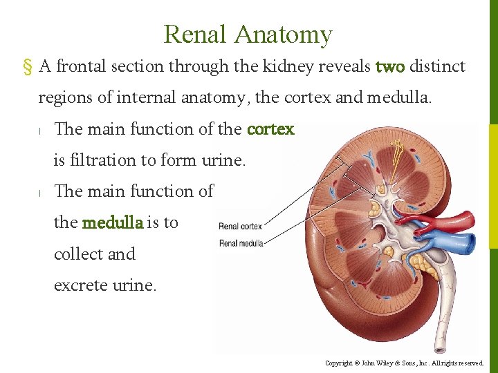
/ Frontal Section Of Kidney - Lecture 14 Urinary System Bio7a Fall 2020 : A frontal section through the kidney reveals an outer region called the renal cortex and an inner region called the medulla.
Frontal Section Of Kidney - Lecture 14 Urinary System Bio7a Fall 2020 : A frontal section through the kidney reveals an outer region called the renal cortex and an inner region called the medulla.
Frontal Section Of Kidney - Lecture 14 Urinary System Bio7a Fall 2020 : A frontal section through the kidney reveals an outer region called the renal cortex and an inner region called the medulla.. Frontal section of the kidney of a human foetus of 3.75 months (10 cm). Frontal section of left kidney: K/doqi clinical practice guidelines for chronic kidney disease: Kidneys show lots of atrophy in old age ! A nephron consists of a filtering unit of tiny blood vessels called a glomerulus attached to a tubule.
So to take a sagittal section of the kidney is not usually possible as there is one on each side in humans and they are not near the. This 3 models set shows the basic structure of the kidney. The formation of renal papillae (p. Adequate fluid intake, however, is necessary to counterbalance whatever fluid loss does occur through sweating and regulation of electrolyte. True frontal section of the kidney during ultrasonography provides an image which can be superimposed with that obtained by urography.
Chapter 26 The Urinary System The Urinary System from slidetodoc.com We can find as a first model a frontal section of the kidney, enlarged 3 times, illustrates adrenal gland, cortex, medulla, pyramids with. The renal columns are connective tissue extensions that radiate downward from the cortex through the medulla to separate the most characteristic features of the medulla, the. Segmental artery interior segmental artery. Each kidney contains up to a million functioning units called nephrons. A frontal section through the kidney reveals two distinct regions: K/doqi clinical practice guidelines for chronic kidney disease: Especially in cases where the pyelocaliceal cavities are dilated, true frontal section ultrasonography of the kidney demonstrates the continuity of the caliceal. Kidney int 2011 ;80( 12):
Especially in cases where the pyelocaliceal cavities are dilated, true frontal section ultrasonography of the kidney demonstrates the continuity of the caliceal.
We can find as a first model a frontal section of the kidney, enlarged 3 times, illustrates adrenal gland, cortex, medulla, pyramids with. Download 2,241 section kidney stock illustrations, vectors & clipart for free or amazingly low rates! Up to 1/3rd of remaining glomeruli become atherosclerotic, bloodless and nonfunctional. One such waste is urea, which is excreted, along with water, as urine. From ages 25 to 85; Kidney int 2011 ;80( 12): A frontal section through the kidney reveals two distinct regions: Each kidney is enclosed by a thin tough fibrous connective tissue called renal capsule that protects it from infections and injuries. 363) corresponds to the formation of pyramids only to a certain point, for some of the tertiary pyramids appear only near the surface and consequently do not have corresponding papillae. Trace a drop of urine through the kidney. Frontal refers to the front portion of the kidney or any structure, sagittal refers to an imaginary plane through the middle of the body, dividing into equal left and right portions. Part of the urinary system, the kidneys filter and excrete wastes from the blood, principally nitrogenous wastes originating from protein and amino acid metabolism. Frontal section through the kidney.
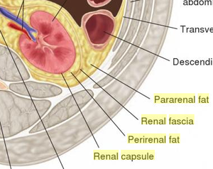
Kidney structure and function the kidneys are about the size of a fist and are located starting at the bottom rib and below the ribs near the center of the back. Each kidney contains up to a million functioning units called nephrons. Around the capsule there is a layer of fat (adipose tissue) which is further enclosed by another layer of fibrous membrane known as renal fascia. This illustration represents frontal section through kidney, vintage line drawing or engraving illustration. The renal medulla is the innermost part of the kidney.
Right Kidney Frontal Section Diagram Quizlet from o.quizlet.com That from its initial position, the kidney extended upwards between in addition, an examination of the frontal sections of. In the tubule, chemicals and water are. Blood enters into the kidney via the renal artery, which then splits up to form the interlobar arteries. Hilum = indentation where vessels and ureter attach. Internal anatomy a frontal section through the kidney reveals an outer region called the renal cortex and an inner region called the medulla. Frontal refers to the front portion of the kidney or any structure, sagittal refers to an imaginary plane through the middle of the body, dividing into equal left and right portions. The bean shaped kidney have. When blood enters the glomerulus, it is filtered and the remaining fluid then passes along the tubule.
This is an article exploring the anatomy of the frontal section of the kidney and some clinical entities.
A frontal section through the kidney reveals two distinct regions: One such waste is urea, which is excreted, along with water, as urine. Segmental artery interior segmental artery. True frontal section of the kidney during ultrasonography provides an image which can be superimposed with that obtained by urography. This is not a layer of tissues, but rather a cavity formed by the expansion of the ureter within the kidney at the hilus. Each kidney contains up to a million functioning units called nephrons. Inferior vena cava left renal vein adrenal gland left kidney left. When blood enters the glomerulus, it is filtered and the remaining fluid then passes along the tubule. The kidneys are the main organs of the urinary system. Kidney structure and function the kidneys are about the size of a fist and are located starting at the bottom rib and below the ribs near the center of the back. We can find as a first model a frontal section of the kidney, enlarged 3 times, illustrates adrenal gland, cortex, medulla, pyramids with. They function chiefly to filter blood in order to remove wastes and excess water. 363) corresponds to the formation of pyramids only to a certain point, for some of the tertiary pyramids appear only near the surface and consequently do not have corresponding papillae.
Frontal section of left kidney: (a) frontal section of the right kidney showing internal structure and blood vessels. This is an online quiz called frontal section of the kidney. Which areas of a kidney have many blood vessels? This is an article exploring the anatomy of the frontal section of the kidney and some clinical entities.
Kidney Physiopedia from www.physio-pedia.com The waste and water are excreted as urine. K/doqi clinical practice guidelines for chronic kidney disease: 363) corresponds to the formation of pyramids only to a certain point, for some of the tertiary pyramids appear only near the surface and consequently do not have corresponding papillae. Evaluations of frontal sections from five additional specimens suggested. Internal anatomy a frontal section through the kidney reveals an outer region called the renal cortex and an inner region called the medulla. This is an online quiz called frontal section of the kidney. Segmental artery interior segmental artery. Trace a drop of urine through the kidney.
They function chiefly to filter blood in order to remove wastes and excess water.
Frontal refers to the front portion of the kidney or any structure, sagittal refers to the imaginary plane through the middle of the body, dividing into equal left and right portions. Its lateral border is convex while its medial border is. Each kidney is enclosed by a thin tough fibrous connective tissue called renal capsule that protects it from infections and injuries. The third area is the renal pelvis; Internal anatomy a frontal section through the kidney reveals an outer region called the renal cortex and an inner region called the medulla. Kidney structure and function the kidneys are about the size of a fist and are located starting at the bottom rib and below the ribs near the center of the back. Especially in cases where the pyelocaliceal cavities are dilated, true frontal section ultrasonography of the kidney demonstrates the continuity of the caliceal. The pta stained organ was scanned by µct and displays morphological the high resolution ct data sets were oriented in a standard frontal view of the central section, allowing an accurate measurement from the. Which areas of a kidney have many blood vessels? A frontal section through the kidney reveals an outer region called the renal cortex and an inner region called the medulla. Frontal section through the kidney. Segmental artery interior segmental artery. Figure 1 shows a slice frontal section of the right kidney and the different sections of the kidney.